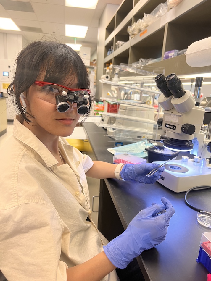
High schoolers Isabella Montoya and Carlos Covarrubias have come to see science as a kind of art. They’ve prepared slides, applied stains, and captured their own stunning images of mouse tissues.
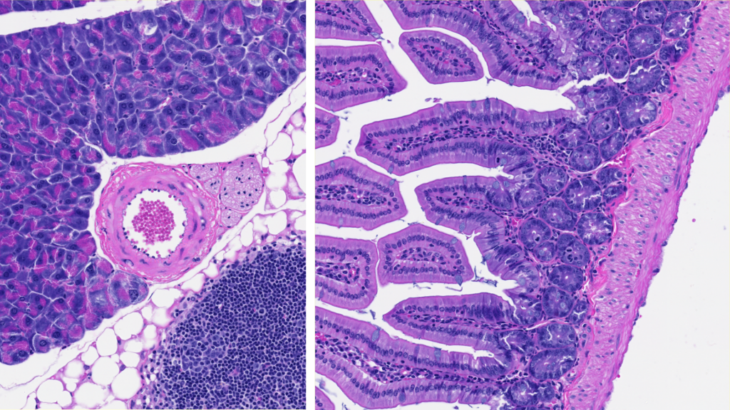
“The most fascinating tissue I have seen under the slide scanner would be the lungs,” says Covarrubias. “Mainly because when fitted with an H&E stain, lung tissue produces a display of colors that resemble art.”
The two are students at High Tech High North County, a charter school in San Marcos, California. Both have a passion for science but never expected to be entrusted with supporting actual immunological studies as high schoolers. Yet as summer interns at La Jolla Institute for Immunology (LJI), Montoya and Covarrubias got to work and learn alongside the highly skilled scientists in the LJI Microscopy and Histopathology Cores.
“I expected to just be following people around to see what they were doing—but we got to do the work too,” says Montoya.
Microscopy and histopathology are key for understanding what goes wrong in the body and how immune cells can help. In microscopy, researchers can illuminate samples and zoom in to spot important structures and even fluorescent tags in tissue samples. Histopathologists also examine tissue samples via microscopy, and they often work with special stains (such as an H&E stain that turns tissues pink and purple) to highlight how cells and pathogens interact.
At LJI, the core staff in the two disciplines work very closely with each other, and their work guides research across the Institute.
Montoya and Covarrubias got a crash course in this work. “There were a lot of acronyms and unfamiliar topics at first, but I learned over time,” says Covarrubias.
Montoya loved getting to explore the features of different tissues. “My favorite part has been prepping tissues to go in the slides and learning about the different stains, like how there are certain stains that only some parts of a sample can absorb,” says Montoya. “I took more than 200 photos of each tissue. These images are so pretty to look at, but it’s not about the pretty colors. I learned about how each stain highlights different parts of the tissue.”
“I want to come back to LJI and see if I can study and work here,” she adds.