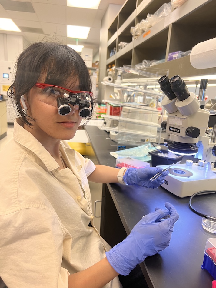
William B. Kiosses, Ph.D., likes the adage “Image is everything.” As a cell biologist, anatomist, and microscopist, he knows how imaging can drive science forward with vision and imaginable clarity.
“To understand cellular science, you kind of have to see it,” says Kiosses, who serves as a La Jolla Institute for Immunology (LJI) Program Projects Core Microscopy Director and Senior Staff Scientist. “It is much easier to explain the behavior of cells when you can see their intracellular and extracellular dynamic world around and in them through moments captured or filmed.”
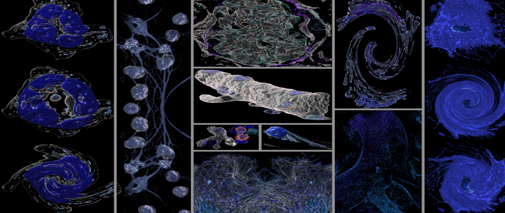
Kiosses’ work is all about collaboration. Scientists at LJI come to him with big questions about how to image or track the complex intracellular world of the cell or how to capture an elusive disease process. Kiosses’ can then lead the imaging effort and show his fellow scientists the microscopy tools and techniques that will shed light on their work.
Kiosses uses a range of enhanced and super-resolution imaging systems analysis tools and techniques to design microscopy-based cell biology assays in static and real time for the scientists he collaborates with.
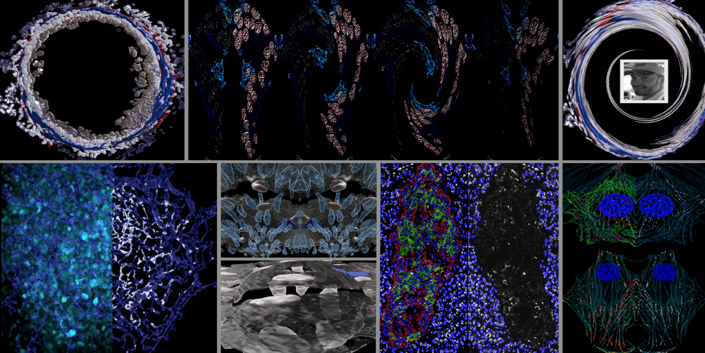
“Microscopy helps create an ideal canvas to illustrate how the many types of immune cells work together in different types of tissues and in disease,” he says. “I can capture spatially, even mechanistically, how the endothelial cells that line the vessels of our cardiovascular system respond to injury, with cohorts of immune cells working synergistically nearby—how they heal wounds or how their inability to heal could potentially pose complications down the road.”
The fruits of these labors are evident in the collection of journal covers credited to Kiosses. “I’ve been lucky and fortunate that I have one or two of those,” says Kiosses.
In truth, Kiosses’ work has been featured on many scientific journal covers. Some journals choose to feature studies with the greatest scientific impact. Other journals pick studies with the most interesting visuals. Kiosses has the skills to highlight both the impact of the work and the imagery.
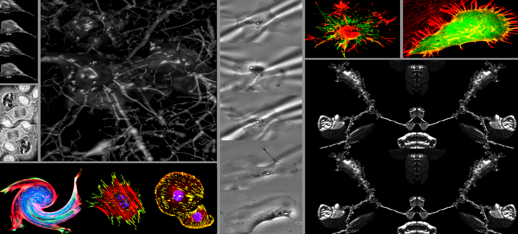
Once a study comes out or a journal cover is published, Kiosses likes to step back and look at the scientific images as an artistic medium. He puts together eye-catching collages of scientific images, highlighting aspects he finds the most striking. “It’s science reimagined as a form of art,” he says.
Kiosses has a hard time narrowing down the list of artists who have influenced him. “I have always admired the visual arts, from the scientific drawings of Da Vinci to the abstractness of Dali, which is reflected in some of my compositions. However, at the same time I’ve always been drawn to the scientific art works of Hooke, Cajal, Boveri, Vesalius, and Netter. I truly appreciate all art,” says Kiosses. “That’s why it’s hard for me to say there is one person or one individual or even one era that actually resonates most.”
“After all, image is everything. The canvas of the microscopic world is never blank, and it’s always evolving,” he adds.