Team: Mehdi Benkahla
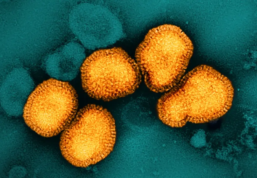
It is thought that an interaction between predisposing genes and environmental factors, such as viral infections, may trigger T1D. Several viruses, mainly from the Enterovirus genus have been considered as potential causal agents for human T1D. Infection studies have been done in mouse models, β cell lines or isolated islets. Here we’ll explore a new model, recently developed by Panzer et al., 2020. This new model consists of alive pancreatic slices obtained from organ donors. We will use them to model enteroviral infections in the human pancreas from non-diabetic donors. In this initial step, we will define a basic protocol that causes infection without severe cytotoxicity, based on the rationale that enterovirus infections in the T1D pancreas appear low grade and possibly chronic, and that severe acute infections have not been observed even in donors with recent onset T1D, whether in nPOD or EADB autopsy cases. We will infect pancreas slices with fully competent CBV3-eGFP. The slices will be washed to eliminate any residual virus. The infections will be monitored as long as the slices remain viable and achieve a low-grade infection as determined by co-staining for VP1 and insulin. Our CVB3-eGFP virus can be used with live cell imaging to determine if β cells, α cells, δ cells, acinar cells, or islet-resident macrophages are productively infected with CVB3 by staining with cell-specific fluorochrome-conjugated antibodies. In collaboration with the microscopy core at La Jolla Institute for Immunology, the von Herrath lab will detect the eGFP-marked virus with high precision. The human pancreas slices will be fixed and co-stained for insulin, glucagon, and somatostatin. We will determine whether the eGFP accumulates in the exocrine or endocrine tissue and if the β cells are more susceptible to the enteroviral infection and hence harboring more eGFP.