Despite microscopy’s long history—the first microscopes date back more than 400 years—new innovations push the imits of what we can see. Yet, increased magnification by itself doesn’t mean much. “Blowing up a tiny picture doesn’t reveal more details, there is a physical limit of how much we can see,” says Zbigniew Mikulski, Ph.D., who directs LJI’s Microscopy and Histology Core. “However, there are some smart ways in which we can break through that barrier. The goal of all technical advances is to improve resolution, be gentler with our samples, penetrate tissues deeper, and in some cases to do so in living animals.”
Today’s microscopes are marvels of engineering—and mind bogglingly expensive—but it still takes human ingenuity to bring never before seen details into view. Dr. Mikulski and his team of microscopy experts stay up-to-date on the latest technology, advise, write software, and even experiment with 3D-printers to help LJI researchers expand their gaze into uncharted depths of living tissues to better understand how the immune system works. The following are some examples of pioneering images that had a powerful impact on LJI scientists’ work.
Skipping the beat
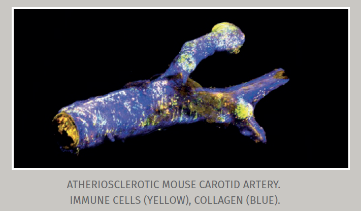
As part of her graduate studies, Sara McArdle, Ph.D., explored the role of immune cells in atherosclerosis, the buildup of fatty substances and cholesterol deposits on the inside of arteries. Specifically, she wanted to use intravital microscopy to track immune cells inside the carotid artery in living mice, but the ebb and flow of blood pulsing through the vessel would inevitably thwart any efforts to record clear videos.
To compensate for the heartbeat, Dr. McArdle designed and custom-built a clever system that links the frequency of the animal’s pulse with the acquisition speed of a 2-photon microscope. This allows the microscope to skip the heart’s beat and acquire a series of stable images.“Right now, nobody in the world does this better than the LJI imaging core,” says Dr. Mikulski. After some computa-tional fine-tuning of the collected data, Dr. McArdle was able to watch as a specific type of immune cell orchestrates the inflammatory assault on the artery wall.
Reflecting the invisible
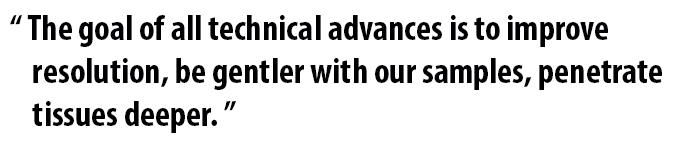
When Gooyoung Seo, Ph.D., wanted to study T cells within intestinal villi, the finger-like projections extending into the interior of the small intestine, she used confocal reflection microscopy (CRM) to trace the villis’ surface relative to the T cells. While most modern microscopes rely on fluorescent tags to identify and visualize molecules, CRM—a technique often used in material sciences—uses polarized light to form images of unlabeled objects placed close to a glass surface.
Much to Dr. Seo’s surprise, CRM revealed that in the ileum, the section of the small intestine next to the appendix, the villi were covered with tiny fibers. Further experiments confirmed that these fibers were segmented filamentous bacteria (SFB), elusive members of the gut microbiome. Dr. Seo is now following up on her unexpected discovery, hoping to learn whether there is a connection between intestinal T cells, the presence or absence of SFBs, and inflammatory bowel disease.
A picture worth more than a thousand words
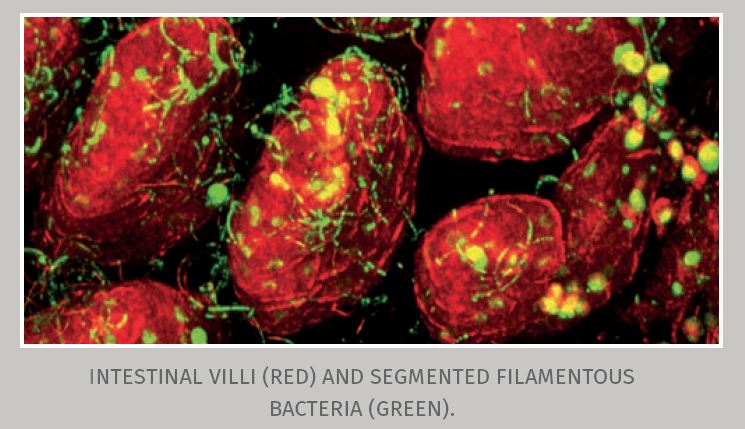
Catie Crosby, Ph.D., is interested in how a special class of immune cells known as invariant natural killer T cells (iNKT) function in the lung during infection with Streptococcus pneumoniae. The bacterium is part of our body’s natural ecosystem but when the immune system is weakened it can turn into a formidable foe. Not only is it the most common cause of community-acquired pneumonia and meningitis in the elderly, it can also manifest itself in myriad different ways.
When Dr. Crosby submitted a grant application to the American Lung Association, she proposed an ambitious project: a combination of intravital microscopy and flow cytometry to study why iNKT cells are so critical for protection against S. pneumonia with the ultimate goal of harnessing the potential of these cells for human therapeutics. Submitting visual proof that LJI’s imaging core was more than up to the task of probing lung tissue of living, breathing mice convinced the selection committee to award her the grant.


